|
If you like things simple and prefer simple explanations - GO AWAY!
GO AWAY NOW! This page will melt your head. The more we see of that hand of creation the more we wonder - but but why????? Couldn't it at least be something alphabetic? Or made
of blocks, wide at the bottom and narrow at the top? Does it always have to be so efficient?
For those of you into flagellation and wrapping your thighs in a cinch of barbed wire, OK, you can substitute this.
In the anatomy section we got a taste in the section called THIGH.
What that section did not mention is that linkages are two way affairs modulating physics by way of muscle action and sensation. Yes - sensation.
Let us consider a VERY common consequence of linkages when we ask the most simple of questions:
Name the knee flexors.
OK. The hamstrings medial (semitendinosus, semimembranosus) and lateral (biceps femoris, both heads). Mmmm, oooh the sartorius. There. Done.
Nope. Add to that list the medial and lateral gastrocnemius (ohhh, of course) and the plantaris, popliteus. Duh, yes, of course those too. Now we are done. No?
No. Add to the list tibialis anterior (WHAT???) , the foot intrinsics and plantar fascia (HEY, THAT AIN'T RIGHT!!!!),
the calcaneus, the toes and iliacus, psoas major and minor and the rectus femoris.
The rectus femoris is a knee extensor! NO WAY!
Way. It is also a hip flexor and with the toe trapped (cleats on the ground) hip flexion passively flexes the knee. Indeed, far and away most frequent source of knee
flexion is seen in ordinary walking where the so-called knee flexors do none of it. The psoas and hip flexors initiate hip flexion which drives linked knee flexion. If the leg is turned out a bit, like a
soccer player, then add the adductors to the knee flexor list.
I HATE THIS. I'm feeling weak.
Here is a typical easy link set up. Read the details in the anatomy section.
The deep blue is the femur. The two light blue sides are the patella-tibia on one side and the pelvis on the other. Red arrows are the hip flexion/knee extension
muscles. The green are the hamstrings (hip extension/knee flexion.
With the right proportion of knee and hip motion these very large muscles need not change length at all. Movements which utilize this feature will be very controlled
and proportional. The single joint muscles shift the balance of the parallelogram being one way or the other.
Go eat something high in carbs.
Brief mind reprieve: From a sensory perspective, the two joint muscles cannot
'know' which shifted position they are in. Only the single joint muscles have certain feed back. When the computation gets difficult the single joint muscles get
disproportionate reliance for knowing what is what. For that reason (figured out in reverse from what we witnessed as effects) we avoid lengthening one joint muscles
to the degree that is possible - within reason. Changing length and at the same time changing the PERCEPTION of length, position, and acceleration is often too much
and results in a longer rehab. Particularly in the blind kids, where they lack a firm spatial reference to reset the rest, we may opt to improve them in smaller steps -
make them better but not go for perfect - too far - too much to relearn - too long a set back.
OK, that thought had a time to percolate. Let us look at it again. The following is a big file (mpeg 4 '.m4v') so give it some time. Depending on your software you
might be able to use the Quicktime© slidy bar (scrub bar) at the bottom to step through.
So how does the hamstring machine which has the feet free cause plantar fascia pain? If you point your feet to render the gastrocnemius muscles limp, it does not.
Otherwise the linkage puts a big unaccustomed repetitive load through a small structure.
Aha! But there is more. The linkage is NOT just mechanical. In a sense, in a linkage sense, the tibialis anterior shares in the information so transmitted by this
links in a chain. This may be small in many cases, but in situations where the main players are not reporting back properly or even not at all (spina bifida, for example
) this L4 (lumbar level 4) innervated muscle may be the only foot posture sensation. Meaning well, somebody fuses the ankle joint or renders it totally immobile. A
sensory loss follows by way of lost SENSORY linkage.
In cases where sensory systems are not fully working, braces that allow dampened motion (we call them flimsy, or maybe springy would have been a better term
although that implies that the mechanical return is more than what we have in mind), how about less rigid? Less rigid braces have better results by way of first not
knocking off sensory links; and second by the deformation creating a differential pressure on skin which creates a new sensory interpretive path. Free hinges lack
this second advantage. Elastic straps to counter hinges may act in a way similar to flimsy braces. If the braced skin is included in the sensory loss area then little is gained from any of this.
Here is a mental test, a thinking hurdle, to use when you are about to pronounce just what a certain bone or muscle does: Use a trapeze catcher as your FIRST
image. Then wander from that point outward. Remember the hand of the creator is not driven by common sense, but rather uncommon sense. In medicine, and
science generally, the sentence that begins "It stands to reason..." has a high probability of being wrong.
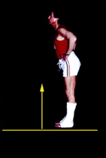
Here is another very common one that we have talked about in many places: The ground reaction linkage.
  With a stiff ankle (by cast, by brace, or by spastic or rigid muscle over push) the foot as a whole becomes the leverage on the shin - driving it backward. Either the knee bends
backward (back knee or recurvatum in Latin) or the hip flexes. Both solve getting the center of the body mass over the center of the foot. Over and over (and over...) we hear this
called truncal weakness. It is NOT! Toe walking avoids ground reaction and thus circumvents both the jack knife posture mistakenly called truncal weakness and also
circumvents recurvatum. Toe walking is not harmful if you have the coordination and balance to handle it. Getting the 'heels to the ground' is often VERY harmful if this is what it produces. With a stiff ankle (by cast, by brace, or by spastic or rigid muscle over push) the foot as a whole becomes the leverage on the shin - driving it backward. Either the knee bends
backward (back knee or recurvatum in Latin) or the hip flexes. Both solve getting the center of the body mass over the center of the foot. Over and over (and over...) we hear this
called truncal weakness. It is NOT! Toe walking avoids ground reaction and thus circumvents both the jack knife posture mistakenly called truncal weakness and also
circumvents recurvatum. Toe walking is not harmful if you have the coordination and balance to handle it. Getting the 'heels to the ground' is often VERY harmful if this is what it produces.
The workings of the fingers is an explosion of complex linkages. Fortunately, we are not going there. Let the hand surgeons explain that mess (mental mess, it works
great as you are aware).
There is another section dealing with linkages from another angle:
MORE about LINKAGE
|