|
|
|
|
|
||||||||||||||||||||||||||
 |
||||||||||||||||||||||||||
|
The Ankle The ankle is a region and the ankle is a joint, but the joint isn't the same region as the region. Huh? What folks call the ankle (singular) is actually two distinctly different joints. What they call ankle motion is actually motion of both those joints. |
|
The joint named 'ankle' is made up of the tibia, the fibula, and the talus. Below the talus is another joint called the 'below the talus joint' (not everybody is a poet) or in another language the subtalar joint. That joint between the talus and calcaneus is intricately inseparable from the action of the midfoot talo navicular joint (and other stuff as well). We'll make sense of this. Here's a question.
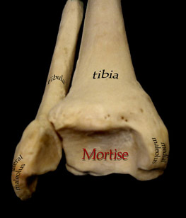
There are three articulations in the mortise: The fibular lateral malleolar, the tibial medial malleolar, and the tibial dome portion. The cylindrical top of the "ankle bone" rolls like a cylinder within that mortise centered by two tough ligaments - one each from the tips of the malleoli.
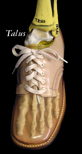
That's the talus - in white - the bone we call the ankle bone. The growth plates (epiphyses) of the fibula and tibia are shown. The talus had a very special application in days gone by. Dice were carved from them, hence the term talisman. That is kind of poetic for there is clearly a major function of the talus to deal with chaos and the unanticipated. When we walk we must anticipate what will happen next so that we can send ahead orders (nerve delivery) as to what to do next. Familiarity and experience make that possible. But the ground can be soft or hard, yielding or firm, sticky or slippery and it may be uneven or look even to collapse unevenly when trod upon. There just isn't time to send back the data and get a correction down to the foot. Sooooo, the ankle region has a built in toggle. The walking orders go out expecting a certain landing which is reflected in the relative tensions of the muscles that control the foot. To the degree that reality does not match expectation, the talus allows the foot to tilt, shift, or lean to accommodate. The talus has a forward projection with a rounded smooth front - the talar head. That projection is in line with the great toe. The Great toe and the metatarsal and bones back to the talus are called the 1st ray. The talus sits on the calcaneus, or heel bone which is in line with the 5th toe. That is called the 5th ray or often the lateral column. The talus can slide forward on the calcaneus or back tethered by ligaments including one called the spring ligament.
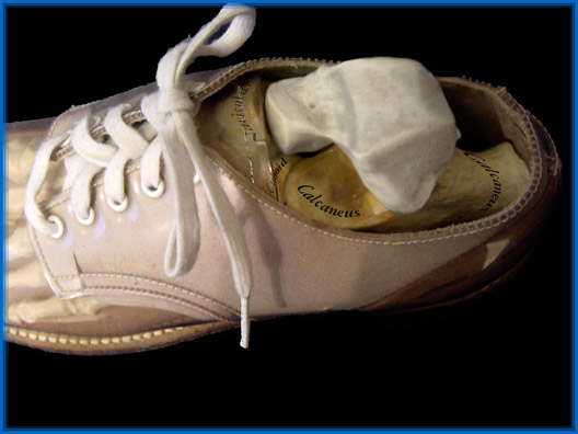
This shows the relationship to the calcaneus better. The calcaneus is not just a "heel" bone, as it is rather long as you can see. It supports the talus by an arm that juts out medially. The joint beneath the talus is called the subtalar joint. Much of the "ankle" motion that is seen is really subtalar motion. The ankle goes up and down in the mortise. The rest is subtalar motion. There are all sorts of descriptions of how the subtalar joint moves, some very biomechanical and mathematical. But they just don't get it. The joint does not "move" but rather allows motion within constraints of set tensions. The big tension setter is the Achilles tendon which delivers the pull of the calf muscles. The Achilles attaches to the heel at the very back surface. That becomes a fixed point. That point is the point of a cone lying on its side. The foot moves in a near conic trace around that tethered point as the inner workings of talus, calcaneus, navicular and the rest adapt to ground forces and Achilles resistance. One can think of the head of the talus as a ball in a ball and socket joint. The navicular and calcaneus provide the socket. However, the talus can piston as well by displacing the navicular forward sliding on the calcaneus.
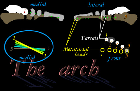
The side roll of the calcaneus itself beneath the talus lowers it closer to the ground as the foot points outward (tracing that cone undersurface outward). Thus the first ray is one which from the toe region points steeply upward toward the head of the talus. The fifth ray points more horizontally toward the calcaneus. The 2nd, 3rd, and 4th take intermediate positions between the outer two columns. Like string art, that creates the arch. From this we see that flat feet point out and high arched feet point more inward as the arch does not "fall" as if a bridge were collapsing. The arch is lost to sideways roll of the calcaneus lowering the talus and with it the first ray. To create an arch, the correct thing is to make the heel vertical, by rolling back under the talus. See also: (muscle version) (heel pain) (movie) Here's another movie=>
The talus points to the navicular which leads to the great toe line. That line already on the floor at the toe is falling at the midfoot so that the rest of it comes down to the floor. So to recreate the arch, just pushing up on the arch area misses the whole deal. By correcting heel valgus (that fall medially) the arch space creates itself with nothing there. Push up there without righting the calcaneus all you get is pain. It won't go far that way. So you see it is really the subtalar joint where the big action takes place - between the talus and the calcaneus. While elongating the lateral side of the foot causes the navicular to displace medially and lift the arch, that is indirect. If the calcaneal valgus is ignored that trick can and will fail - sooner or later. You need to ask WHY did the calcaneus fall over sideways dropping the talus medially? In many kids it is just like a lazy eye. The medial muscle whose job it is to hold up the medial side is just plain off circuit. You watch thee struggle when you ask them to walk on the lateral border of their feet. Might as well ask them to wiggle their ears. The lack of participation of the medial support muscles is the cause. Can't ignore it. In spastics there may be active down ward puller forces. Yes, in ligamentous laxity it may crumble as well. What we know about intrinsic structural foot shape integrity is also a bit weird - taking away structure from the medial side does not collapse the foot. The collapse happens when you remove the ligaments from around the calcaneal cuboid area especially near where an extension of the tibialis posterior tendon shoots a bit of tendon. The point? Often missed is that mildly flat feet can be way more painful than some very nasty looking dead flat feet. Why? Because the mild ones are not FLAT FEET. They are ordinary feet being wrenched by tight gastroc-Achilles and mother earth into a forced tilt to accommodate the missing calf muscle range. In other words tight Achilles can make stuff hurt. The Achilles will rupture before it complains. So other stuff becomes painful and gets attention. If the quarterback is getting bloody, look to the defect in the line not his nasal septum. An AFO molded with the calcaneus in valgus will SUSTAIN the flat foot and arches added at the arch area just plain hurt. Correction requires that the ANKLE be able to turn outward and move laterally relative to the heel center.
|
|
|
||||||||||||||||||||
 The fibula is a bone which is often borrowed to build
other parts that need bone stock. We can do rather well with the bulk of the shaft missing. We need the top bit for the ligaments which attach there. We definitely need the lower part because that completes the ankle mortise.
The fibula is a bone which is often borrowed to build
other parts that need bone stock. We can do rather well with the bulk of the shaft missing. We need the top bit for the ligaments which attach there. We definitely need the lower part because that completes the ankle mortise.